
Periodontology Pictures 1
Test Summary
0 of 45 Questions completed
Questions:
Information
You have already completed the test before. Hence you can not start it again.
Test is loading…
You must sign in or sign up to start the test.
You must first complete the following:
Results
Results
0 of 45 Questions answered correctly
Your time:
Time has elapsed
You have reached 0 of 0 point(s), (0)
Earned Point(s): 0 of 0, (0)
0 Essay(s) Pending (Possible Point(s): 0)
Categories
- Not categorized 0%
- 1
- 2
- 3
- 4
- 5
- 6
- 7
- 8
- 9
- 10
- 11
- 12
- 13
- 14
- 15
- 16
- 17
- 18
- 19
- 20
- 21
- 22
- 23
- 24
- 25
- 26
- 27
- 28
- 29
- 30
- 31
- 32
- 33
- 34
- 35
- 36
- 37
- 38
- 39
- 40
- 41
- 42
- 43
- 44
- 45
- Current
- Review
- Answered
- Correct
- Incorrect
-
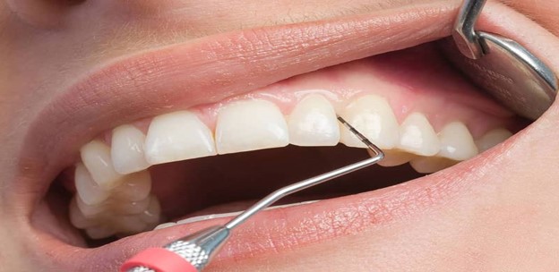
Question 1 of 45
1. Question
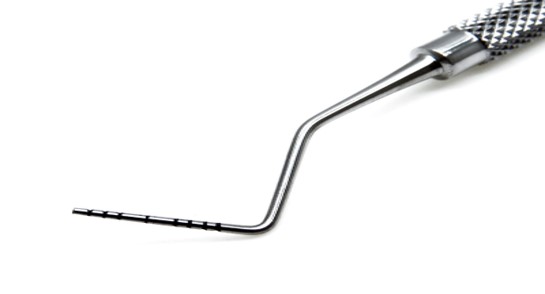
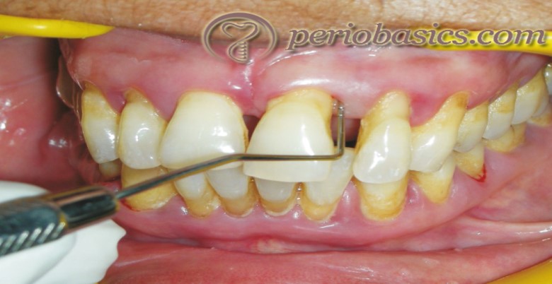
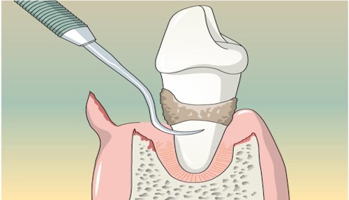
What is the purpose of the instrument shown in the image?
CorrectIncorrect -
Question 2 of 45
2. Question
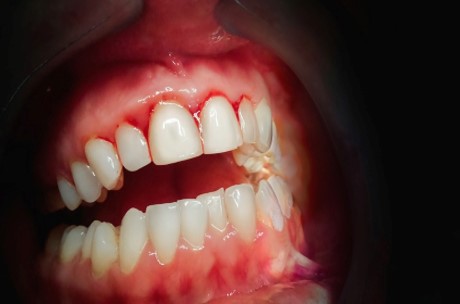
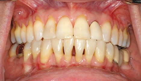
This patient has localized deep periodontal pockets around a molar, and the gingiva appears erythematous and swollen. What is the likely diagnosis?
CorrectIncorrect -
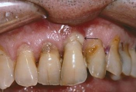
Question 3 of 45
3. Question
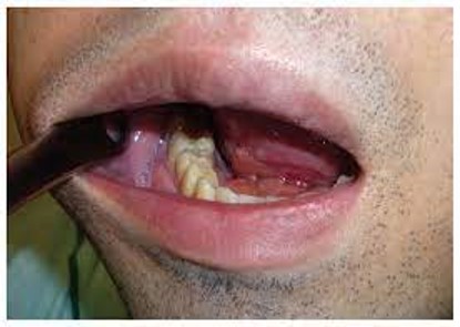
The procedure shown in the image is commonly used to treat:
CorrectIncorrect -
Question 4 of 45
4. Question
The above image shows a dental probe reaching the bottom of the gingival sulcus with no attachment loss. What is the likely diagnosis?
CorrectIncorrect -
Question 5 of 45
5. Question
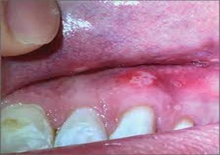
This image shows localized redness, swelling, and a spontaneous gingival abscess. What is the likely cause?
CorrectIncorrect -
Question 6 of 45
6. Question
This patient presents with vertical bone loss and deep periodontal pockets on both sides of a tooth. What term describes this type of bone loss?
CorrectIncorrect -
Question 7 of 45
7. Question
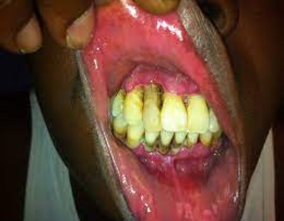
What is the most likely diagnosis for this inflamed, swollen gingiva with purulent discharge?
CorrectIncorrect -
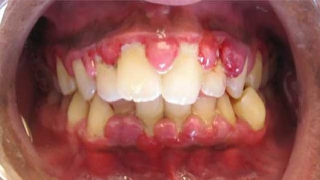
Question 8 of 45
8. Question
This patient exhibits sudden onset severe pain, fever, and a swelling filled with pus in the gingival tissue. What is the likely diagnosis?
CorrectIncorrect -
Question 9 of 45
9. Question
This dental probe encounters resistance, preventing it from reaching the base of the gingival sulcus. What does this suggest?
CorrectIncorrect -
Question 10 of 45
10. Question
This image shows swollen, erythematous gingiva with prominent strawberry-like appearance. Which viral infection is most likely associated with this clinical presentation?
CorrectIncorrect -
Question 11 of 45
11. Question
In the image, there is widespread gingival inflammation with interdental papillae necrosis. What condition does this represent?
CorrectIncorrect -
Question 12 of 45
12. Question
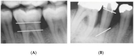
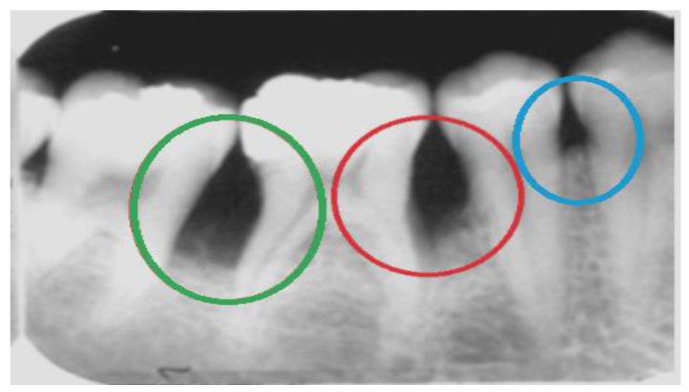
Image of a dental radiograph showing a horizontal bone loss pattern around a tooth. What type of periodontal pocket is commonly associated with this pattern?
CorrectIncorrect -
Question 13 of 45
13. Question
Observe the gingival involvement in this image. What systemic condition is most likely associated with this finding?
CorrectIncorrect -
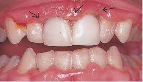
Question 14 of 45
14. Question
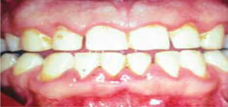
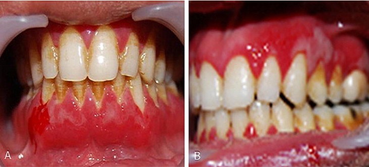
A patient presents with swollen, red, and bleeding gums. Dental examination reveals deep periodontal pockets. What is the likely diagnosis?
CorrectIncorrect -
Question 15 of 45
15. Question
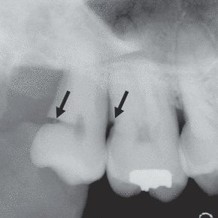
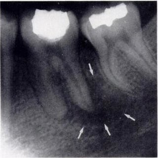
Image of a dental radiograph shows a notch-like defect in the interdental alveolar bone. What is the likely cause of this pattern?
CorrectIncorrect -
Question 16 of 45
16. Question
What is the most concerning feature depicted in this image for a patient with acute gingival infection?
CorrectIncorrect -
Question 17 of 45
17. Question
An image shows diffuse swelling of the sublingual and submandibular spaces. What is the most probable diagnosis?
CorrectIncorrect -
Question 18 of 45
18. Question
The above image displays localized bone loss around the furcation area of a multi-rooted tooth. What is the likely term for this condition?
CorrectIncorrect -
Question 19 of 45
19. Question
Identify the characteristic feature in this image suggesting a specific acute gingival condition in children.
CorrectIncorrect -
Question 20 of 45
20. Question
The image shows a radiograph revealing a radiolucency at the apex of a tooth root. What is the likely diagnosis?
CorrectIncorrect -
Question 21 of 45
21. Question
A patient exhibiting generalized vertical bone loss with a “floating tooth” appearance. What term describes this advanced condition?
CorrectIncorrect -
Question 22 of 45
22. Question
This image shows gingival involvement in a patient with a specific blood dyscrasia. What is the most likely diagnosis?
CorrectIncorrect -
Question 23 of 45
23. Question
The above patient presents with painful, swollen, and bleeding gums associated with erupting wisdom teeth. What condition is most likely responsible?
CorrectIncorrect -
Question 24 of 45
24. Question
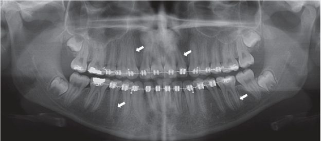
In this image, there is radiographic evidence of a widened periodontal ligament space. What is the likely cause of this finding?
CorrectIncorrect -
Question 25 of 45
25. Question
What is the primary function of the gingiva?
CorrectIncorrect -
Question 26 of 45
26. Question
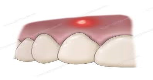
This image displays localized, fluctuant swelling at the gingival margin with pus drainage. What is the likely diagnosis?
CorrectIncorrect -
Question 27 of 45
27. Question
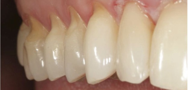
A patient exhibits a characteristic “V”-shaped notch at the cervical area of teeth. What is the likely etiology of this pattern?
CorrectIncorrect -
Question 28 of 45
28. Question
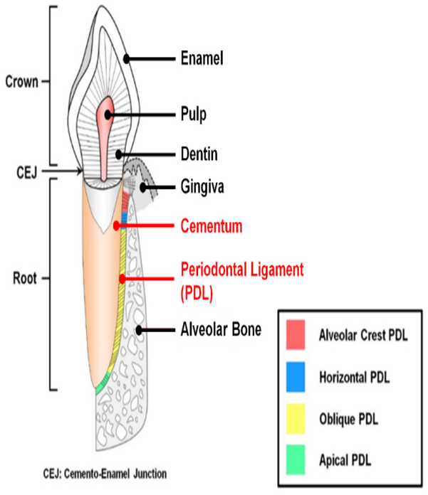
What is the role of the periodontal ligament?
CorrectIncorrect -
Question 29 of 45
29. Question
This patient presents with multiple painful ulcers and a grayish pseudomembrane on the gingiva. What is the likely diagnosis?
CorrectIncorrect -
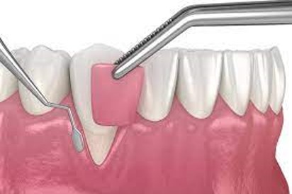
Question 30 of 45
30. Question
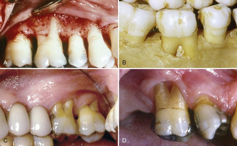
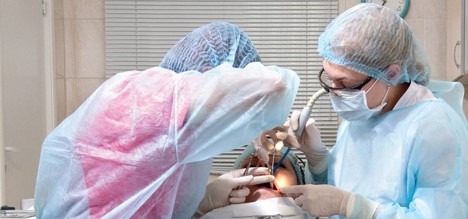
The image shows a surgical procedure where a portion of the gingiva and underlying tissues is elevated to access the underlying bone. What type of surgery is being performed?
CorrectIncorrect -
Question 31 of 45
31. Question
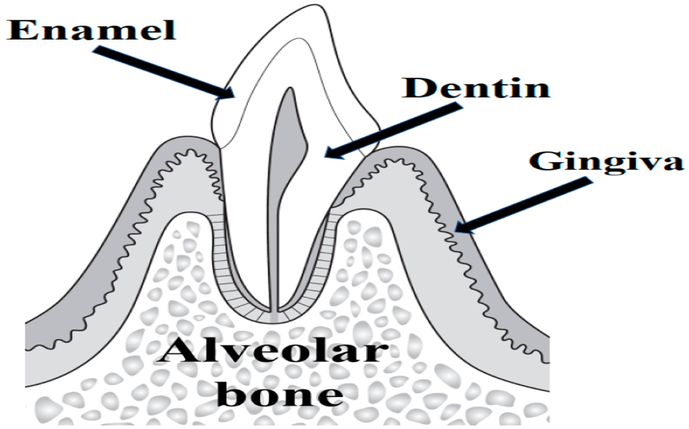
Which part of the tooth-supporting structures houses the tooth sockets?
CorrectIncorrect -
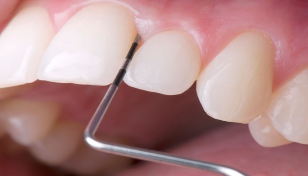
Question 32 of 45
32. Question
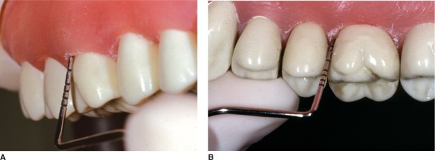
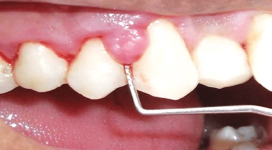
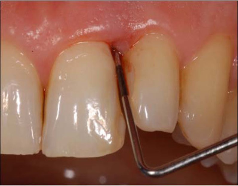
This image shows a dental probe inserted into a gingival sulcus, measuring a depth greater than 3mm. What is the likely diagnosis?
CorrectIncorrect -
Question 33 of 45
33. Question
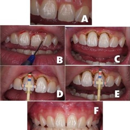
In the above image, a dentist is creating an incision that follows the contour of the gingival margin. What type of incision is being made?
CorrectIncorrect -
Question 34 of 45
34. Question
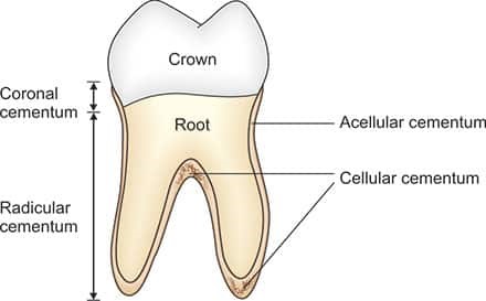
What is the function of cementum in tooth anatomy?
CorrectIncorrect -
Question 35 of 45
35. Question
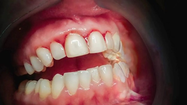
In the image, the probe reveals bleeding upon gentle probing with no visible attachment loss. What condition does this likely represent?
CorrectIncorrect -
Question 36 of 45
36. Question
A patient undergoing surgery to enhance the width and thickness of the attached gingiva. What is the term for this mucogingival surgical procedure?
CorrectIncorrect -
Question 37 of 45
37. Question
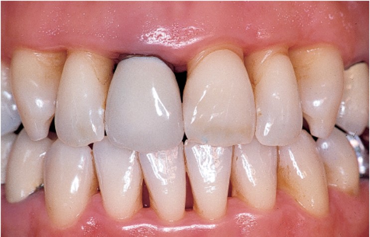
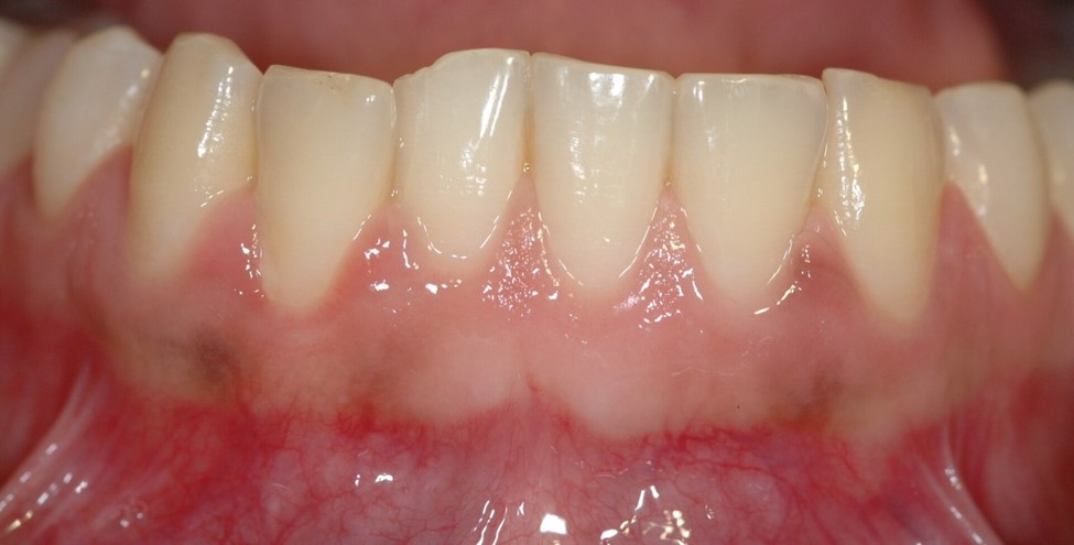
The condition depicted in the image is:
CorrectIncorrect -
Question 38 of 45
38. Question
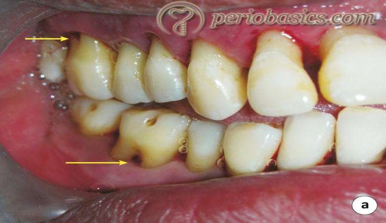
A dental radiograph showing bone loss around a tooth with a deep periodontal pocket. What is the most probable diagnosis?
CorrectIncorrect -
Question 39 of 45
39. Question
A dentist performs a procedure to remove excess gum tissue to expose more of the tooth crown. What is this procedure called?
CorrectIncorrect -
Question 40 of 45
40. Question
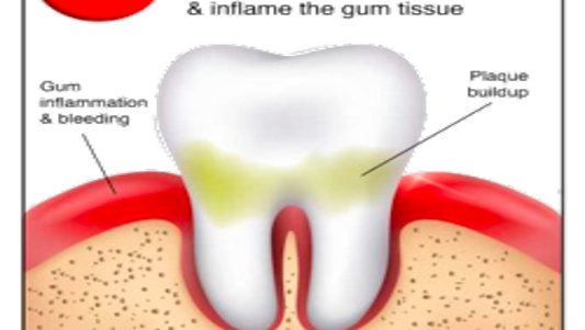
What is the primary cause of gingivitis?
CorrectIncorrect -
Question 41 of 45
41. Question
The above image displays a dental probe extending beyond the apical extent of the gingival sulcus. What term is appropriate for this condition?
CorrectIncorrect -
Question 42 of 45
42. Question
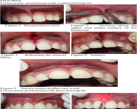
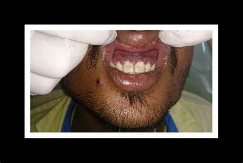
In the image, a dentist performs a procedure to release a tight labial frenulum. What is the term for this surgical procedure?
CorrectIncorrect -
Question 43 of 45
43. Question
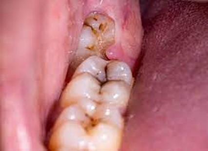
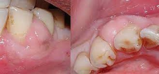
The condition shown in the image is indicative of:
CorrectIncorrect -
Question 44 of 45
44. Question
Identify the characteristic clinical feature in this image suggestive of a specific acute gingival infection.
CorrectIncorrect -
Question 45 of 45
45. Question
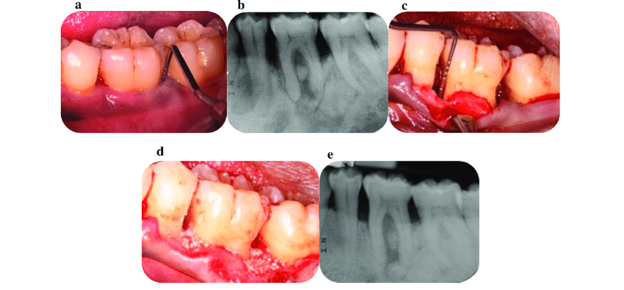
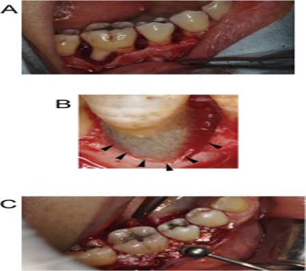
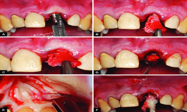
Identify the surgical procedure depicted in this image, where a full-thickness gingival flap is reflected to expose the underlying bone.
CorrectIncorrect